


Fluid Mosaic Model Structure, Components, Functions, and Diagram
How do the components of a cell membrane work together? Membranes are described by the fluid mosaic model and this helps us understand how cell membranes are able to hold their shape but still permits many of the components within to move around and interact with each other. They are not hard, immovable walls but flexible structures composed mostly of phospholipids, proteins and other molecules that allow cells to communicate, transport and control their interior.
S. J. Singer and Garth L. Nicolson introduced this membrane model in 1972. Nicolson that this model changed the way scientists think about biological membranes. It illustrates a phospholipid bilayer with a variety of proteins, cholesterol and carbohydrate-containing molecules.
Once you understand the fluid mosaic model, you can better understand: Why substances enter and exit the cell membrane Cell-cell recognition Membrane proteins’ role in carrying out the needs of the cell.
This guide presents the model’s definition, structure, parts, functions, scientific evidence, examples, and contemporary modifications-with a diagram with labels and tables for easier visualization.
What Is the Fluid Mosaic Model?
The fluid mosaic model is a scientific hypothesis which explains the structure of the plasma membrane, the cell’s outermost membrane.
It defines a membrane as a phospholipid bilayer with proteins and other molecules that are free to move within the membrane creating a flexible yet ordered construct.
The fluid mosaic model explains how phospholipids, proteins, and other molecules form a dynamic yet organized cell membrane, providing a foundation for understanding its structure and functions. For a more detailed explanation, explore this fluid mosaic model definition and its role in cell membrane structure
The phrase “fluid mosaic” refers to two important traits:
- Fluid: Many membrane lipids and proteins can move laterally within the membrane.
- Mosaic: Different molecular components are distributed throughout the membrane, forming a varied arrangement.
The model explains how a cell membrane can remain intact while supporting transport, signaling, recognition, and changes in cell shape.
Who Proposed the Fluid Mosaic Model?
Biological membrane structures were described by S. J. Singer and Garth L. Nicolson in their paper “The Fluid Mosaic Model in Science” in February 1972. They suggested that the membranes are lipid bilayers with embedded and dispersed proteins that are spatially arranged in relation to their associated functions or chemistry.
This model succeeded previous accounts where membrane proteins were viewed essentially as continuous fill covering the membrane bilayer.
Why Is It Called a Mosaic?
A mosaic is an arrangement of different pieces that combine to form a larger structure.
In a cell membrane, these molecular components include:
- Phospholipids that form the bilayer.
- Integral and peripheral proteins.
- Cholesterol in many animal membranes.
- Glycoproteins and glycolipids containing carbohydrate chains.
Their different structures and functions create the membrane’s mosaic organization.
Fluid Mosaic Model Illustration: A Visual Guide to Cell-Membrane Structure
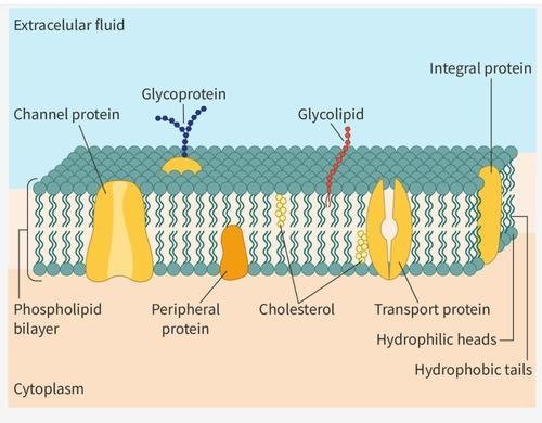
Figure 1 of 2. This illustration shows the cell membrane’s phospholipid bilayer, embedded proteins, and carbohydrate-bearing components.
Start by looking at the diagram of a fluid mosaic model. Locate the two phospholipid layers in the diagram. The two hydrophilic heads are rounded and face the watery environment inside or outside of the cell. The two hydrophobic heads are pointed to the inside of the membrane.
After that, locate the proteins that are embedded in or attached to the bilayer. Cholesterol molecules are found in the spaces between phospholipids in animal membranes, while carbohydrate chains linked to lipids or proteins tend to extend out from the plasma membrane’s outer surface.
This illustration shows a simplified cross section. In practice, membranes are rather more complex three dimensional chemically varied structures that are arranged differently depending on the type of cell.
Structure of the Fluid Mosaic Model
The model explains how lipids, proteins, and carbohydrates are organized within the plasma membrane. Each component contributes to membrane stability, flexibility, communication, or transport.
1. Phospholipid Bilayer
A double layer of phospholipids provides the membrane’s fundamental structural foundation. Each phospholipid contains a water-attracting head and two water-repelling fatty acid tails.
These molecules are amphipathic, meaning they contain both hydrophilic and hydrophobic regions.
In an aqueous environment, phospholipids arrange themselves into two layers:
- Hydrophilic heads face the extracellular fluid and the cytoplasm.
- Hydrophobic tails point inward, away from surrounding water.
This arrangement creates a barrier that helps regulate the movement of substances across the membrane.
2. Integral and Peripheral Proteins
Membrane proteins are responsible for a wide range of vital functions including transport, signal transfer, enzyme activity and structural interaction.
Integral membrane proteins are embedded within the phospholipid layer, and some extend across it. Some span the whole membrane; others are inserted only in part of it.
Peripheral proteins are either attached to the membrane surface or interact with the other membrane proteins but are not embedded through the hydrophobic core.
A channel protein, for instance, creates a hole that certain ions or molecules can pass through.
3. Cholesterol
Cholesterol, which is found in most animal cell membranes, is another lipid. It behaves like a buffer, wedged between phospholipids and affecting their fluidity.
This helps the membrane to stay flexible as its temperature changes.
4. Glycoproteins and Glycolipids
Glycoproteins are proteins with attached carbohydrate chains. Glycolipids consist of lipid molecules bonded to short carbohydrate chains. They play a role in cell recognition, signaling, and interactions with the environment.
For instance, surface carbohydrates allow cells to identify molecular identities and are involved in interactions regulating tissue architecture and immune recognition.

Components of the Fluid Mosaic Model and Their Functions
The following table summarizes the main components of the fluid mosaic model and explains how their structures relate to their biological roles.
| Component | Location or structure | Main function |
| Phospholipids | Form the two-layer membrane framework | Provide the basic barrier and membrane structure |
| Integral proteins | Embedded within the bilayer | Transport, signaling, and molecular interactions |
| Peripheral proteins | Associated with membrane surfaces | Enzymatic activity and structural support |
| Cholesterol | Between phospholipids in many animal membranes | Helps regulate membrane fluidity |
| Glycoproteins | Proteins with attached carbohydrate chains | Cell recognition and communication |
| Glycolipids | Lipids with attached carbohydrate chains | Cell recognition and membrane interactions |
The relative proportions of these components vary between cell types and organelles. For example, membranes specialized for transport or energy-related processes may have different protein and lipid compositions.
Channel Proteins and Carrier Proteins
Channel proteins create pathways that only certain molecules can pass through membranes. They can be controlled by signals or they can be opened whenever needed.
Carrier proteins attach to specific molecules and change shape to assist in their transport across the membrane.
Crucially, not all carrier proteins use ATP Some (pulls itself along a concentration gradient) moves passively, others actively transport substances.
Why Is the Cell Membrane Fluid?
Membrane fluidity refers to the ability of membrane molecules to move and reorganize within the lipid bilayer.
This movement is important because the membrane must support molecular interactions without behaving like a completely rigid structure.
Lateral Movement
Most phospholipids and membrane proteins have the ability to move laterally within the same membrane. This is known as lateral diffusion.
This enables the molecules to rearrange and to interact with adjacent molecules.
But not all membrane protein can freely diffuse. The membrane protein’s lateral diffusion is limited by interactions with other proteins, the cytoskeleton, or other structures.
Other Types of Molecular Movement
The molecules within the membrane can also rotate in the membrane.
The other type of movement, known as ‘flip-flop’ or ‘transverse diffusion’ involves the molecule moving from one phospholipid layer to the next. This type of movement is usually far less frequent for phospholipids than lateral diffusion and may require an enzyme.
Factors That Affect Membrane Fluidity
Several factors influence the physical behavior of the membrane.
| Factor | General effect |
| Higher temperature | Usually increases molecular movement |
| Lower temperature | Usually decreases molecular movement |
| Unsaturated fatty acid tails | Tend to increase fluidity |
| Longer fatty acid tails | Tend to reduce fluidity |
| Cholesterol | Moderates temperature-related changes in fluidity |
The effect of cholesterol depends on temperature and membrane composition. It should not be described simply as a substance that always increases fluidity.
Functions and Importance of the Fluid Mosaic Model
The fluid mosaic model helps explain how membrane structure supports several essential cellular functions.
1. Selective Permeability
The plasma membrane controls what comes in and goes out.
Small nonpolar molecules can cross the lipid bilayer more easily than charged particles or large polar molecules.
Other substances require membrane transport proteins.
This ensures that cells can maintain the ideal conditions required for biological function.
2. Transport of Molecules
Membrane proteins support the movement of nutrients, ions, and other substances.
Three important transport processes include:
- Simple diffusion: Movement down a concentration gradient without direct energy input.
- Facilitated diffusion: Passive movement through a channel or carrier protein.
- Active transport: Movement that requires energy, directly or indirectly, to move substances against an electrochemical gradient.
Osmosis, the movement of water across a selectively permeable membrane, is another important process.
3. Cell Communication
Receptors on the membrane sense cues in the environment.
For instance, a signal molecule could attach to a receptor protein and cause activity inside a cell. Cell signalling enables cells to respond to hormones, growth factors and other signals from their environment.
4. Cell Recognition
Carbohydrate-containing molecules on the outer membrane surface help cells recognize and interact with one another.
This is important in tissue development, immune responses, and cellular organization.
5. Flexibility and Shape Changes
The fluid nature of the membrane enables a variety of cell shape changes that include membrane fusion, vesicular transport, and other activities.
Red blood cells, for instance, need to squeeze through tiny capillaries. Their organization of the membrane and structural proteins keeps the cells flexible.
History and Development of the Fluid Mosaic Model
Prior to the fluid mosaic model, other theories were proposed about the arrangements of lipids and proteins within cell membranes.
An alternative theory that had significant influence in the past was the Davson-Danielli sandwich model of the membrane which had a phospholipid bilayer with protein sheets on both surfaces.
Microscopy and biochemical work refuted this view by showing that membrane structure was more complex than this.
In 1972, Singer and Nicolson suggested that proteins may not form a complete continuous sheath outside the membrane but were rather positioned within the lipid bilayer.
By including the hypothesis of lateral movement of molecules within the membrane plane, and the effects of molecular properties and interactions on organization, their model was developed.
Fluid Mosaic Model vs. Sandwich Model
| Feature | Davson–Danielli sandwich model | Fluid mosaic model |
| Protein arrangement | Protein layers coating the bilayer | Proteins embedded in or associated with the bilayer |
| Membrane dynamics | Limited representation of molecular movement | Emphasizes lateral mobility |
| Organization | Relatively uniform layered structure | Diverse molecular components |
| Historical significance | Earlier membrane explanation | Proposed in 1972 and subsequently refined |
The fluid mosaic model provided a more useful framework for understanding membrane proteins, molecular mobility, and membrane function.
Evidence Supporting the Fluid Mosaic Model
The fluid mosaic model is supported by experimental observations that reveal membrane organization and molecular movement.
Freeze-Fracture Electron Microscopy
Freeze-fracture microscopy allows researchers to examine internal surfaces of biological membranes. Observations of particles associated with membrane fracture faces provided evidence consistent with proteins embedded within the lipid bilayer.
Cell Fusion Experiments
Researchers used distinct labels on membrane proteins in classic cell fusion experiments to track their movement.
The labeled proteins, after two cells had fused, spread over the cell membrane, confirming that membrane proteins are capable of lateral movement.
Fluorescence Recovery After Photobleaching (FRAP)
FRAP is a method that is used to study molecular mobility.
A researcher with a fluorescently tagged membrane molecule can bleach out a tiny patch of it with a laser then watch to see if it repopulates as unbleached molecules diffuse into the patch.
Recovery rate and extent inform about the nature of the motion and the number of molecules that are mobile.
These methods, collectively, demonstrate that membranes are indeed fluid but the rate of motion depends upon the characteristics of the interacting molecules.
Modern Understanding and Limitations of the Fluid Mosaic Model
While the fluid mosaic model still forms the basis of the explanation of membrane organization, current studies have developed the original image.
The membrane should not be viewed as a homogeneous sheet in which molecules constantly move freely.
Membrane Domains and Lipid Rafts
A membrane domain is an area of a membrane, which has a different molecular organization and composition than the rest of the membrane.
Lipid raft is described as a specialized membrane domain enriched in specific lipids and proteins. Lipid raft is implicated in several cellular functions including signaling and membrane organization.
Cytoskeletal Interactions
The inside of the cell is made up of the cytoskeleton, a web of structural proteins.
Certain membrane proteins bind to elements of the cytoskeleton either directly or through intermediaries. These interactions are what make it possible for the proteins to control or be affected by diffusion.
Membrane Asymmetry
The inner and outer layers of a biological membrane do not necessarily contain identical lipid compositions.
This asymmetry contributes to membrane properties and cellular functions.
Why the Original Model Has Been Refined
The 2023 review by Nicolson and Ferreira de Mattos describes how later research added membrane domains, cytoskeletal assemblies, extracellular matrix interactions, and more complex molecular organization to the original model.
| Simplified original picture | Modern understanding |
| Membrane components move within a fluid bilayer | Movement may be restricted by molecular interactions |
| Proteins are distributed within a lipid matrix | Proteins may form complexes and specialized domains |
| Membrane represented as relatively uniform | Membrane organization can vary across regions |
| Lipid–protein interactions emphasized | Cytoskeletal and extracellular interactions also matter |
These developments refine the fluid mosaic model rather than making its central principles obsolete.
Real-Life Examples of the Fluid Mosaic Model
Understanding the model becomes easier when its components are connected to actual cellular activities.
Red Blood Cells: Membrane Flexibility
Red blood cells have to navigate the blood vessels – including tiny capillaries.
Their membranes had to undergo large shape changes while still remaining intact. That’s an example of why flexibility and architecture of membranes are important.
Nerve Cells: Ion Channels
Nerve cells rely on membrane proteins that regulate the movement of ions.
Ion channels contribute to electrical signaling by allowing selected ions to cross the membrane under appropriate conditions.
Intestinal Cells: Nutrient Transport
Cells lining the intestine use membrane transport proteins to absorb nutrients.
For example, glucose transport involves specific membrane proteins that help move glucose across cellular membranes.
Immune Cells: Recognition
Membrane-associated molecules help immune cells interact with other cells and recognize molecular features.
Carbohydrate-containing surface molecules contribute to these recognition processes.
Cell Signaling: Membrane Receptors
A receptor protein can bind a signaling molecule outside the cell and initiate a response inside it.
This demonstrates how membrane proteins support communication between a cell and its environment.

Common Misconceptions About the Fluid Mosaic Model
Misconception 1: Every membrane protein moves freely.
Not necessarily. Some proteins are restricted by interactions with other membrane molecules, the cytoskeleton, or extracellular structures.
Misconception 2: The membrane consists only of phospholipids.
The membrane also contains proteins and other molecules, including cholesterol in many animal membranes and carbohydrate-containing components.
Misconception 3: Cholesterol always makes the membrane more fluid.
Cholesterol moderates membrane fluidity, and its effect depends on temperature and membrane composition.
Misconception 4: The fluid mosaic model describes a completely disorganized membrane.
The membrane is dynamic but organized. Molecular interactions and specialized domains influence how its components are distributed and move.
Misconception 5: The model applies only to animal cells.
The model describes fundamental principles of biological membrane organization, although membrane composition differs among organisms, cell types, and organelles.
Frequently Asked Questions
1. What is the fluid mosaic model in simple words?
The fluid mosaic model describes the cell membrane as a flexible double layer of phospholipids with proteins and other molecules embedded in it. Some of these molecules are capable of moving about within the membrane giving it a role in transport, communication and recognition.
2. Who proposed the fluid mosaic model?
S. J. Singer and Garth L. Nicolson introduced the fluid mosaic model for membranes in 1972. This model provided a detailed description of the arrangements of proteins and lipids in biological membranes and their lateral mobility.
3. What are the main components of the fluid mosaic model?
The main components include phospholipids, integral and peripheral proteins, cholesterol in many animal membranes, and carbohydrate-containing molecules such as glycoproteins and glycolipids.
4. Why is the cell membrane called fluid?
It is called fluid because many phospholipids and membrane proteins can move laterally within the membrane. This movement allows the membrane to remain dynamic while maintaining its overall organization.
5. What is the role of cholesterol in the fluid mosaic model?
Cholesterol helps regulate membrane fluidity by moderating the effects of temperature on phospholipid movement. Its influence depends on the membrane’s temperature and composition.
6. What evidence supports the fluid mosaic model?
Evidence includes observations from freeze-fracture electron microscopy, cell fusion experiments demonstrating lateral protein movement, and fluorescence recovery after photobleaching, which measures molecular mobility.
7. Is the fluid mosaic model still accepted today?
Yes. It remains a useful foundation for understanding membrane structure. Modern research has refined it by incorporating membrane domains, cytoskeletal interactions, molecular complexes, and restrictions on movement.
Conclusion
The fluid mosaic model explains how the cell membrane combines flexibility with organized molecular structure.The bilayer of phospholipids offer the primitive structure while, proteins, cholesterol, and the molecules with carbohydrates assist in recognition, transport, and membrane integrity.
While newer studies have shown a more sophisticated membrane patterning than what was depicted by the initial 1972 portrayal, its core concepts still lie at the heart of cell biology.
When students understand the organisation, mobility and interaction of the subcellular components, then they can gain better insight into the properties of cells which enables them to function and adapt to their environment.
When students understand the organization, mobility, and interaction of subcellular components, they gain deeper insight into how cells function and adapt to their environment. For readers interested in expanding their knowledge beyond biology, they can explore more insights into science, technology, and modern innovation through additional educational resources.